Deep Lymph Node Imaging at the Cellular Level IVIM TECHNOLOGY
WHITEPAPER
August 2024 |
|
|
Deep Lymph Node Imaging at the Cellular Level: How Intravital Microscopy Revolutionizes Immune Response Tracking and Enables In Vivo Monitoring of Nanoparticle Delivery in the Lymphatic System |
|
|
Introduction
Lymphatic System
The lymphatic system is critical for initiating immune responses by transporting lymph, which contains immune cells, through lymphatic vessels to lymph nodes. Within these nodes, lymphocytes, macrophages, and antigen-presenting cells (APCs) interact to trigger antigen-specific immune responses. This process is orchestrated by a specialized reticular meshwork and involves the filtration and destruction of particulate antigens by macrophages.
Nanoparticle Drugs in the Lymphatic System
Nanoparticles have emerged as powerful tools for targeting lymph nodes, offering a means to deliver therapeutic agents directly to immune cells such as dendritic cells and T-cells. This targeted delivery optimizes immune-related therapies by ensuring that drugs reach their intended cellular targets within the lymphatic system.
Physiological Barriers to Nanoparticle Delivery to Lymph Nodes
Targeting lymph nodes with nanoparticles presents several challenges:
- Avoidance of Clearance: Nanoparticles must evade the mononuclear phagocyte system (MPS) — comprising macrophages, monocytes, and neutrophils — that removes foreign particles from the bloodstream.
- Size and Surface Charge: Nanoparticle stability and circulation time are influenced by size and surface charge. Smaller particles with neutral or negative charges generally have prolonged circulation.
- Extravasation: Nanoparticles need to pass through the blood vessel endothelium, facilitated by their size and shape.
- Extracellular Matrix Interaction: Upon entering the interstitium, nanoparticles must navigate the extracellular matrix (ECM), which can restrict their movement based on size and surface charge.
- Navigating the ECM: Effective delivery to lymph nodes requires overcoming the ECM's complex protein network, where smaller, neutral particles often show better diffusion.
Strategies for Overcoming Barriers to Nanoparticle Delivery to Lymph Nodes
To address these challenges, several strategies can be employed:
- Design Optimization: Developing small, neutral nanoparticles can enhance their ability to traverse physiological barriers and target lymph nodes effectively.
- Targeted Delivery Systems: Crafting systems that bind to endogenous molecules or immune cells that naturally migrate to lymph nodes can improve targeting.
- Cell-Based Delivery: Attaching nanoparticles to immune cells such as T cells, monocytes, and dendritic cells, which naturally traffic to lymph nodes, can enhance drug delivery through both lymphatic and blood vessels.
Intravital Imaging of Nanoparticle Delivery to Lymph Nodes
Incorporating intravital imaging into your strategy provides real-time insights into how nanoparticles interact with lymph nodes. This approach helps you refine designs and delivery systems based on detailed in vivo observations, ultimately improving the efficacy and safety of nanoparticle-based therapies.
Traditional ex vivo and in vitro methods often fall short in replicating the complex interactions and dynamic processes in living organisms. They cannot fully capture immune cell trafficking or the influence of physiological conditions like pH. Consequently, these methods may provide incomplete or inaccurate data on nanoparticle behavior. In contrast, intravital imaging offers real-time observation of nanoparticles within lymph nodes, providing a comprehensive and accurate view of their biodistribution and interactions with tissues. This technique is crucial for evaluating the true efficacy and safety of nanoparticle-based drug delivery systems in vivo.
|
|
|
Materials & Methods
Nanoparticle Injection in Mice
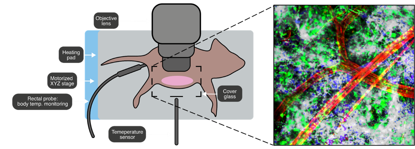
Prox1-GFP transgenic mice, expressing GFP in lymphatic endothelial cells, were utilized for nanoparticle injection and lymph node imaging. We injected 50 µl of nanoparticles subcutaneously into the right flank of each mouse. The control group received 50 µl of saline similarly. To visualize blood vessels, we administered 50 µl of FSD 555-conjugated anti-CD31 antibody (IVITM-991-0003, IVIM Technology, Inc., Korea) intravenously via the tail vein two hours prior to imaging.
Real-time Intravital In Vivo Imaging with Two-Photon Microscopy
Intravital imaging was performed using an IVM-CM (Intravital Confocal and Two-Photon Microscope, IVIM Technology, Inc., Korea). Three days post-injection, the inguinal skin was incised to expose the lymph node, followed by two-photon imaging with a 25X water immersion objective lens. Multi-position z-stack imaging was conducted to capture detailed images. |
|
|
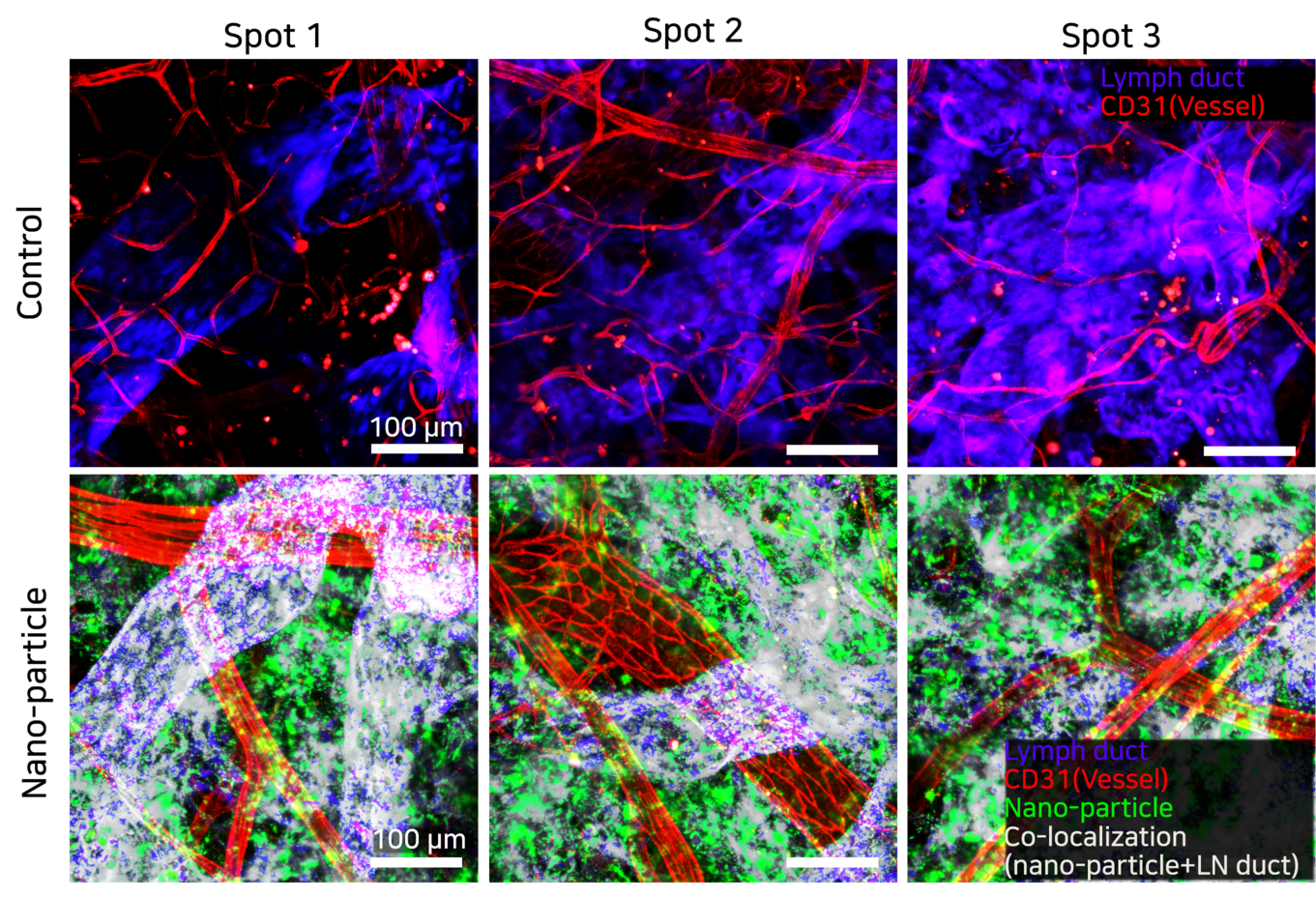
Figure 1. Z-stack images of the control group and nanoparticle group, taken 3 days after saline or nanoparticle injection. Purple/blue is Lymph duct, Red is vessel labeled with CD31, green is nanoparticle, white color refers to co-localization (nanoparticle+ lymph duct) |
|
|
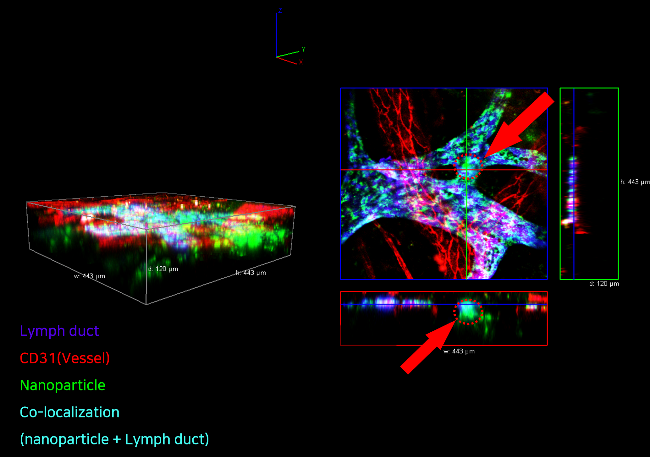
Figure 2. 3D view of nanoparticle distribution in the lymph node, three days after nanoparticle injection. |
|
|
Three days after nanoparticle injection, the nanoparticles were distributed throughout the lymph node, with notable colocalization with the lymphatic duct (Figure 1). Three-dimensional rendering of the images confirmed the spatial distribution of nanoparticles within the lymphatic duct (Figure 2).
Conclusion
Our study examined the delivery and distribution of nanoparticles to the inguinal lymph nodes in live mice, employing intravital imaging for real-time observation. Using Prox1-GFP transgenic mice, we injected nanoparticles subcutaneously and visualized their distribution within the lymph nodes and their colocalization with lymphatic ducts using two-photon microscopy. Intravital imaging enabled us to overcome the limitations of ex vivo and in vitro methods, providing a comprehensive view of the dynamic processes involved in nanoparticle distribution. The 3D imaging confirmed the spatial distribution of nanoparticles within the lymphatic ducts, demonstrating their potential as effective drug delivery systems targeting lymphatic tissues. These findings underscore the importance of real-time intravital imaging in assessing the in vivo behavior of nanoparticle-based therapies, offering valuable insights that could drive future advancements in targeted drug delivery |
|
|
References
- Willard-Mack, C. L. (2006). Normal structure, function, and histology of lymph nodes. Toxicologic Pathology, 34(5), 409-424. https://doi.org/10.1016/j.jsps.2017.10.012
- Rizvi, S. A. A., & Saleh, A. M. (2018). Applications of nanoparticle systems in drug delivery technology. Saudi Pharmaceutical Journal, 26(1), 64-70. https://doi.org/10.1016/j.jsps.2017.10.012
- Trac, N., & Chung, E. J. (2021). Overcoming physiological barriers by nanoparticles for intravenous drug delivery to the lymph nodes. Experimental Biology and Medicine, 246(22), 2358-2371. https://doi.org/10.1177/15353702211010762
- Jeong, S.-H., Jang, J.-H., & Lee, Y.-B. (2021). Oral delivery of topotecan in polymeric nanoparticles: Lymphatic distribution and pharmacokinetics. Journal of Controlled Release, 335, 86-102. https://doi.org/10.1016/j.jconrel.2021.05.017
|
|
|
2024, IVIM Technology, Inc. All rights reserved.
|
|
|
IVIM Technologyinformation@ivimtech.com#A-1305, Hyundai Knowledge Industry Center, 11, Beobwon-ro 11-gil, Songpa-gu, Seoul, 05836, Korea
Tel: +82-2-431-7450Unsubscribe |
|
|
|